We use cookies to enable the functions required for this website, such as login or a shopping cart. You can find more information in our privacy policy.
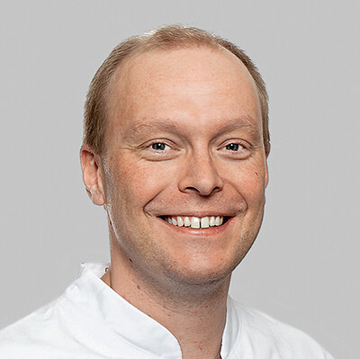
From 1994 to 1999, Prof. Dr. Florian Beuer studied at the Dental School of the Ludwig Maximilian University Munich, Germany, where he gained his license to practice (DDS) in 2000. From 2000 to 2001, he worked as an employed dentist in a private practice. In 2002, he became assistant professor in the Department of Prosthodontics of the Ludwig Maximilian University and made his Dissertation (Dr. med. dent.) in the same year. He became Specialist in Implantology (German Society for Implantology) in 2005. From 2007 to 2008, he worked as a Visiting Professor at the Pacific Dental Institute in Portland, Oregon (Director: John A. Sorensen DMD, PhD). His habilitation (Priv. Doz., PhD) was in 2009 at the LMU. From 2009 to 2015, he was the Vice President of the "Deutsche Gesellschaft für Ästhetische Zahnheilkunde (DGAEZ)". In 2010, he became an Expert in Prosthodontics after a further education at DGPro. Since 2011, Prof. Buer is a board member of the Team AG Ceramics. In the same year, he became the editor of the "Teamwork Journal of Continuing Dental Education". Since 2014, he was full Professor at Ludwig Maximilians University until 2015, when he became the Chairman of the Department of Prosthodontics at the Charité University of Medicine Berlin. He is also a Board Member of the German Society for Implantology (DGI).He gained his Master of Medical Education MME (University of Heidelberg) in 2015.
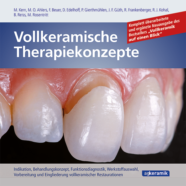
Indikation, Behandlunskonzept, Funktionsdiagnostik, Werkstoffauswahl, Vorbereitung und Eingliederung vollkeramischer Restaurationen
1st completely revised and expanded new Edition 2023 Book 21 x 21 cm, 255 pages Language: German Categories: Prosthodontics, Restorative Dentistry ISBN 978-3-9817012-4-1
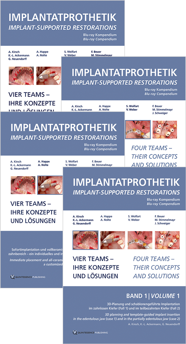
1st Edition 2012 Blu-Ray 4 Blu-ray-discs in box; NTSC/PAL; Laufzeit/Run time: 311 min Language: German, English Categories: Implantology, Prosthodontics, Dental Technology ISBN 978-3-86867-117-9 QP Deutschland
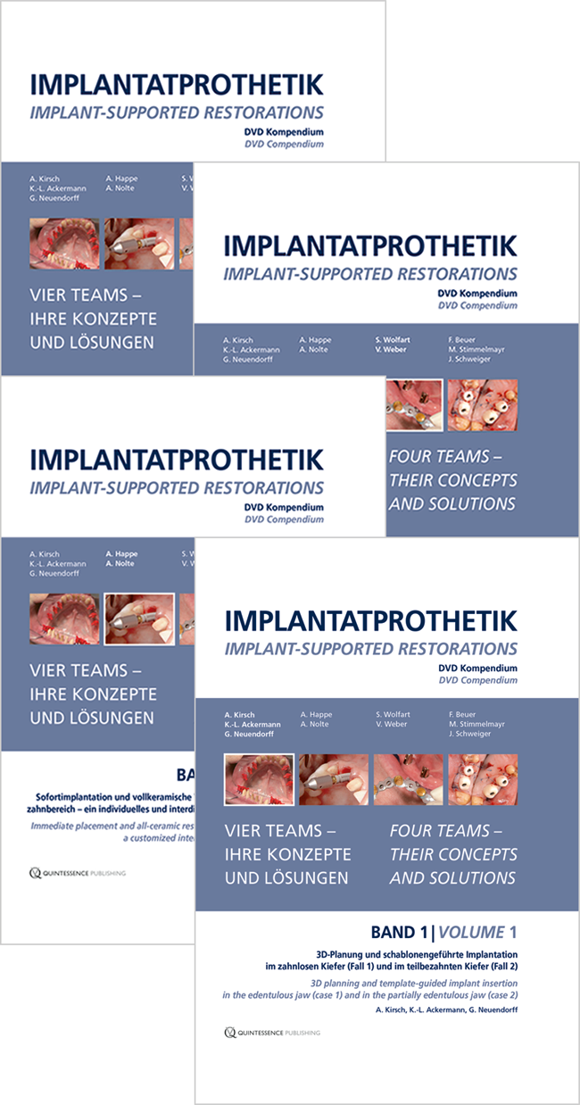
1st Edition 2012 DVD 4 DVDs in Box; NTSC/PAL; Laufzeit/Runtime: 311 min Language: German, English Categories: Implantology, Prosthodontics, Dental Technology Stock No.: C3012 ISBN 978-3-86867-112-4 QP Deutschland
Issue cycle: Quarterly Language: English Impact factor: 1,8 (2023) Categories: Interdisciplinary, General Dentistry Editor-in-chief: Prof. Dr. Florian Beuer MME QP Deutschland
30th EAO Annual Scientific Meeting / 37th DGI Annual Congress
Berlin reloadedSeptember 28, 2023 — September 30, 2023CityCube Berlin, Berlin, Germany
Speakers: Samir Abou-Ayash, Bilal Al-Nawas, Thomas Bernhart, Florian Beuer, Stefan Bienz, Elena Calciolari, Najla Chebib, Andreas Dengel, Vincent Donker, Joke Duyck, Roberto Farina, Gary Finelle, Alberto Fonzar, Tobias Fretwurst, Rudolf Fürhauser, Oscar Gonzalez-Martin, Stefano Gracis, Knut A. Grötz, Christian Hammächer, Lisa J. A. Heitz-Mayfield, Detlef Hildebrand, Norbert Jakse, Jim Janakievski, Tim Joda, Daniel Jönsson, Gregg Kinzer, Vincent G. Kokich, Michael Krimmel, Cecilia Larsson Wexell, Martin Lorenzoni, Georg Mailath-Pokorny, Julia Mailath-Pokorny, Frank Georg Mathers, Gerry McKenna, Henny Meijer, Alberto Monje, Torsten Mundt, Nadja Nänni, David Nisand, Robert Nölken, Nicole Passia, Michael Payer, Christof Pertl, Aušra Ramanauskaitė, Eik Schiegnitz, Martin Schimmel, Ulrike Schulze-Späte, Frank Schwarz, Falk Schwendicke, Robert Stigler, Michael Stimmelmayr, Anette Strunz, Christian Ulm, Stefan Vandeweghe, Kay Vietor, Arjan Vissink, Asaf Wilensky, Stefan Wolfart, Werner Zechner, Anja Zembic, Nicola Zitzmann
European Association for Osseintegration (EAO)
This author's journal articles
International Journal of Computerized Dentistry, Pre-Print
Aim: The adjustment and transfer of a stable occlusion can be a major challenge in prosthetic rehabilitations. The aim of this study was to assess a non-invasive treatment option for complex prosthetic rehabilitations and occlusal analyses using 3D-printed restorations clinically.
Materials and Methods: Eleven patients received a partial or complete rehabilitation with the aid of 3D-printed restorations (n=171). After 12 months of clinical service, all restorations were analyzed using the United States Public Health Service (USPHS) criteria.
Results: The 12-month clinical data revealed that 3D-printed restorations showed a survival rate of 84.4%. Complications occurred mostly regarding the anatomical form (7%) or marginal integrity (6AC%) and were consequently rated “Charlie” or “Delta.” Color stability and color match of 3D-printed restorations were rated “Alpha” in 83% and 73%, respectively, of all restorations. Marginal inflammation was rated “Alpha” in 89% of all restorations. An excellent surface texture and no secondary caries or postoperative sensitivities (100%) were observed.
Conclusions: 3D-printed restorations might be an alternative treatment option for initiating complex prosthetic rehabilitations. Technical complications rarely occurred. Biological complications did not occur at all. The color stability showed promising results after 12 months of clinical service. However, the results should be interpreted with caution. Long-term results with a high number of restorations should be awaited.
Keywords: 3D-printing, additive manufacturing, CAD/CAM, color stability, in vivo, wear behavior
Aim: To report on a novel digital superimposition workflow that enables measuring the supra-crestal peri-implant soft tissue dimensions all along implant treatment and afterwards. Materials and Methods: A preoperative CBCT and intra-oral scans (IOS) are successively taken before surgery, at the end of the healing period, at prosthesis delivery, and over time; they are digitally superposed on a dedicated software. Then, the stereolithography files (STL) of the healing abutment, of the prosthetic abutment and the crown are successively merged into the superposition set of IOSs. Result: The workflow protocol of merging successively the STL of each item into the superposition set of IOSs enables capturing the dimensions of the height and width of the supra-crestal soft tissues, at every level of the healing abutment, the prosthetic abutment and the crown. In addition, it allows measuring the vertical distance that the crown exerts pressure on the gingiva and the thickness of the papillae at every level of the abutment. Conclusion: This novel digital superimposition workflow provides a straightforward method of measuring the vertical and horizontal dimensions of the supra-crestal peri-implant soft tissues, including the papillae, at each stage of the implant treatment process. It allows investigating a certain number of soft tissue variables that were previously inaccessible to clinical research. It should help enhancing our comprehension of the peri-implant soft tissue dynamics.
Keywords: CBCT, clinical research, digital merging, gingival height, gingival width, intra-oral scan, papilla, peri-implant soft tissues
Purpose: A reference method for quantifying contaminations on two-piece abutments manufactured using CAD/CAM has not yet been established. In the present in vitro study, a pixel-based machine learning (ML) method for detecting contamination on customized two-piece abutments was investigated and embedded in a semiautomated quantification pipeline.
Materials and methods: Forty-nine CAD/CAM zirconia abutments were fabricated and bonded to a prefabricated titanium base. All samples were analyzed for contamination by scanning electron microscopy (SEM) imaging followed by pixel-based ML and thresholding (SW) for contamination detection; quantification was performed in the postprocessing pipeline. Wilcoxon signed-rank test and Bland-Altmann plot were applied to compare both methods. The contaminated area fraction was recorded as a percentage.
Results: There was no statistically significant difference between the percentages of contamination areas (median = 0.004) measured with ML (median = 0.008) and with SW (median = 0.012), asymptotic Wilcoxon test: P = 0.22. The Bland-Altmann plot demonstrated a mean difference of -0.006% (95% confidence interval [CI] from -0.011% to 0.0001%) with increased values from a contamination area fraction of > 0.03% for ML.
Conclusion: Both segmentation methods showed comparable results in evaluating surface cleanliness; pixel-based ML is a promising assessment tool for detecting external contaminations on zirconia abutments. Further studies are required to investigate the clinical performance of this tool.
Keywords: computer-aided design, scanning electron microscopy, machine learning, ultrasonics, hygiene, dental implant abutments