Auf unserer Website kommen verschiedene Cookies zum Einsatz: Technisch notwendige Cookies verwenden wir zu dem Zweck, Funktionen wie das Login oder einen Warenkorb zu ermöglichen. Optionale Cookies verwenden wir zu Marketing- und Optimierungszwecken, insbesondere um für Sie relevante und interessante Anzeigen bei den Plattformen von Meta (Facebook, Instagram) zu schalten. Optionale Cookies können Sie ablehnen. Mehr Informationen zur Datenerhebung und -verarbeitung finden Sie in unserer Datenschutzerklärung.
Prof. Dr. Fouad Khoury absolvierte sein Studium der Zahnmedizin an der Saint-Joseph-Universität in Beirut/Libanon und spezialisierte sich an den Universitäten Freiburg und Münster auf Oralchirurgie. An der Universität Münster wurde er zum Oberarzt ernannt und schloss dort auch seine Habilitation ab. Seit 1994 ist er Inhaber einer Professur an der Klinik für Mund-, Kiefer- und Gesichtschirurgie der Universität Münster und Direktor der Privatzahnklinik Schloss Schellenstein, Olsberg, Deutschland. Prof. Khoury ist seit vielen Jahren Gastprofessor an mehreren Universitäten, Vorsitzender der Prüfungskommission für Oralchirurgie und Mitglied des Redaktionsausschusses zahlreicher internationaler Fachzeitschriften. Außerdem war er Vorsitzender der Arbeitsgemeinschaft für Mund-, Kiefer- und Gesichtschirurgie (AGOKI) der Deutschen Gesellschaft für Zahn-, Mund- und Kieferheilkunde (DGZMK) und ist Mitherausgeber des International Journal of Oral Implantology. Er hat mehrere Auszeichnungen erhalten (z. B. den Pioneers in Dentistry Award 2016 der AUB und den William R. Laney Award der American Academy of Osseointegration 2017) und ist Inhaber zahlreicher Patente. Prof. Khoury ist Autor und Herausgeber von vier Lehrbuch-Bestsellern, die in mehr als 10 Sprachen übersetzt wurden.
Referenten: Bilal Al-Nawas, Florian Beuer, Daniel Buser, Vincent Fehmer, Dominik Groß, Knut A. Grötz, Jan-Frederik Güth, Anke Handrock, Arndt Happe, Sönke Harder, Matthias Kern, Marco Rainer Kesting, Fouad Khoury, Torsten Mundt, Sven Reich, Irena Sailer, Eik Schiegnitz, Henning Schliephake, Frank Schwarz, Anton Sculean, Michael Stimmelmayr, Istvan Urban, Kay Vietor, Christian Walter, Stefan Wolfart, Giovanni Zucchelli, Otto Zuhr
Deutsche Gesellschaft für Implantologie im Zahn-, Mund- und Kieferbereich e.V.
ROX 2024 - the Rosa Experience
Bone and Soft Tissue Regeneration22. März 2024 — 23. März 2024São Paulo, Brasilien
Referenten: José Carlos Martins da Rosa, Fouad Khoury, Istvan Urban, Giovanni Zucchelli
71. Jahrestagung der Arbeitsgemeinschaft Oral- und Kieferchirurgie / 42. Jahrestagung des Arbeitskreises für Oralpathologie und Oralmedizin
18. Mai 2023 — 19. Mai 2023Kurhaus Bad Homburg, Bad Homburg v. d. Höhe, Deutschland
Arbeitsgemeinschaft für Oral- und Kieferchirurgie
70. Jahrestagung der Arbeitsgemeinschaft Oral- und Kieferchirurgie / 41. Jahrestagung des Arbeitskreises für Oralpathologie und Oralmedizin
26. Mai 2022 — 27. Mai 2022Kurhaus Bad Homburg, Bad Homburg v. d. Höhe, Deutschland
Arbeitsgemeinschaft für Oral- und Kieferchirurgie
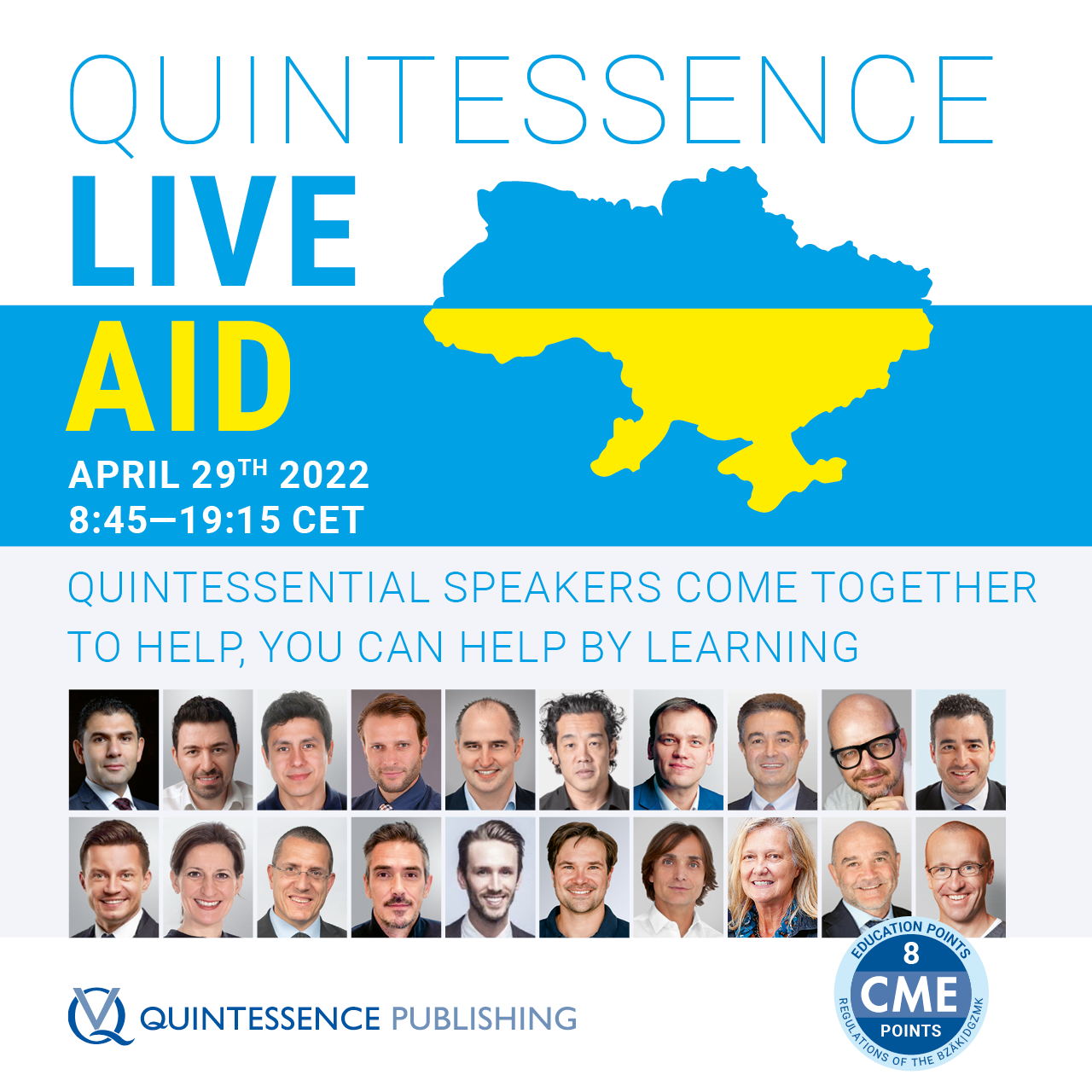
Quintessence Live Aid
Quintessential Speakers from around the world offer a Day of Education that will help those in need29. Apr. 2022Zoom
Referenten: Wael Att, Maxim Belograd, Victor Clavijo, Christian Coachman, Stefan Fickl, Ronaldo Hirata, Fouad Khoury, Tomas Linkevičius, Pascal Magne, Nazariy Mykhaylyuk, José M. Navarro, Irena Sailer, Anton Sculean, Kyle Stanley, Miguel Stanley, Markus Tröltzsch, Istvan Urban, Débora R. Vilaboa, Giovanni Zucchelli, Otto Zuhr
ausgebuchtQuintessenz Verlags-GmbH
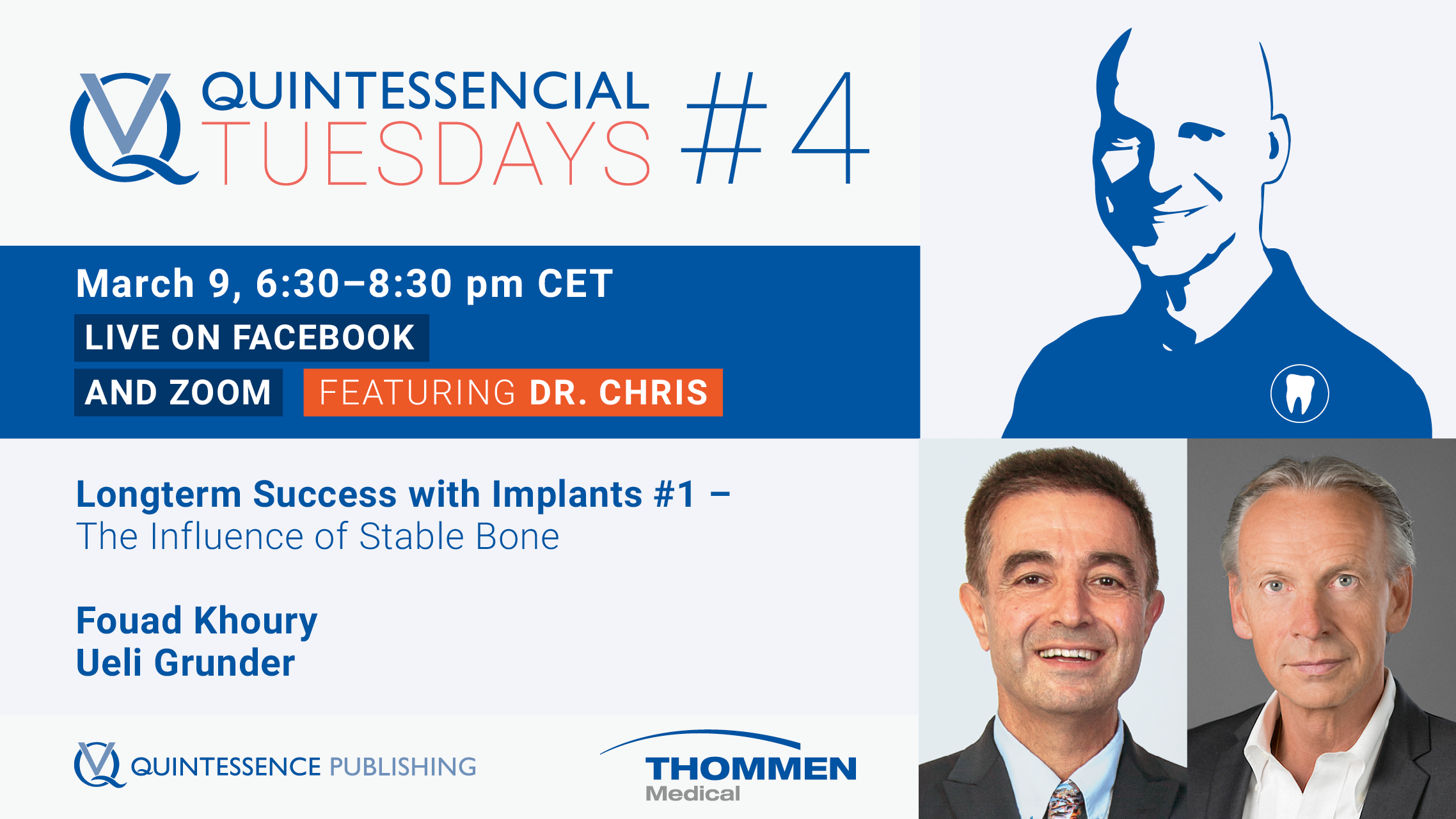
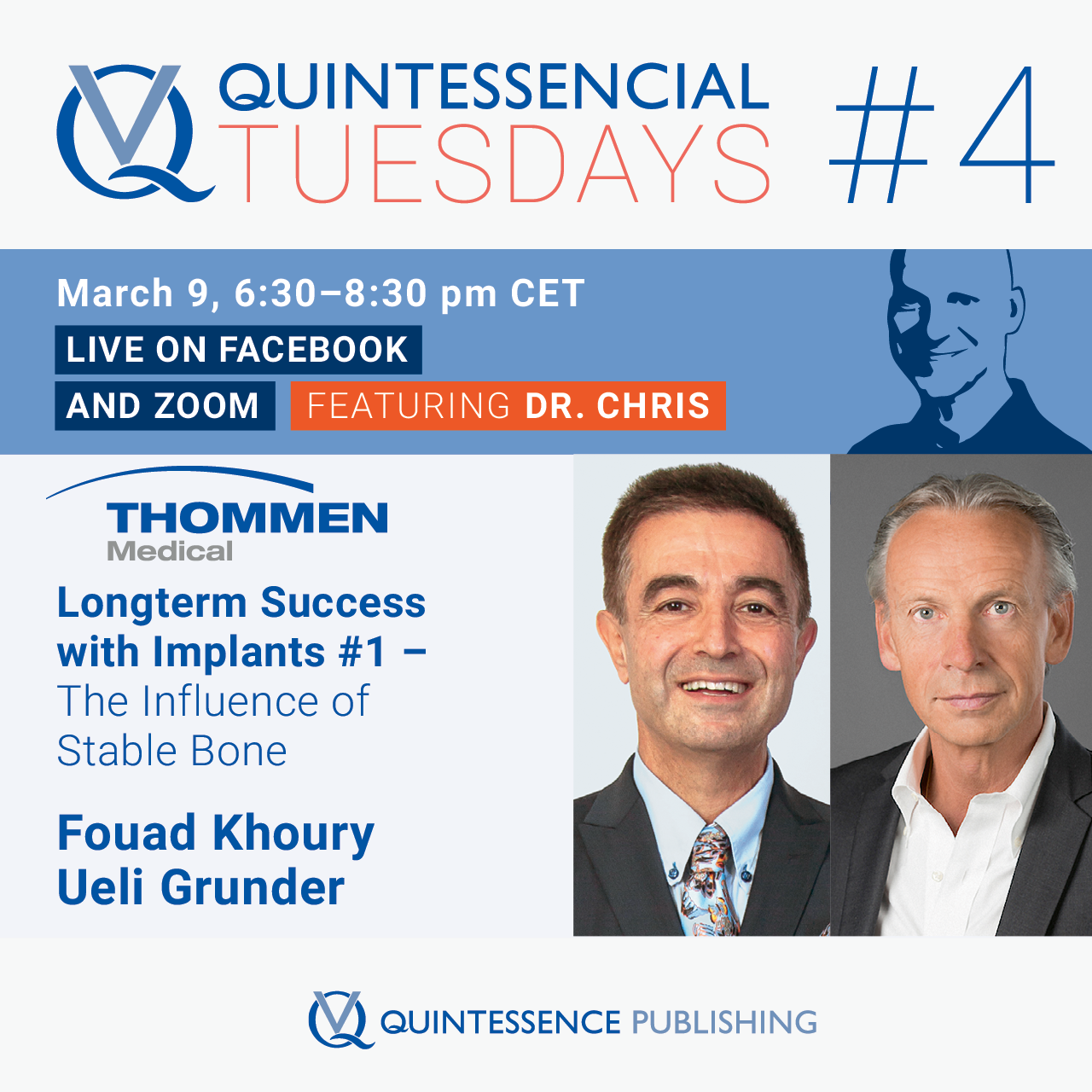
FREE - Quintessencial Tuesday with Dr. Chris #4
Longterm Success with Implants #1 - The Influence of Stable Bone9. März 2021, 18:30 — 20:30 UhrOnline
Referenten: Ueli Grunder, Fouad Khoury, Christopher Köttgen
Quintessenz Verlags-GmbH
Bone Augmentation in Oral Implantology
Advanced Surgical Procedures with Autogenous Bone30. März 2019London, Vereinigtes Königreich von Großbritannien und Nordirland
Quintessence Publishing Co. Ltd. UK
7 Decades of Quintessence Publishing
10. Jan. 2019 — 12. Jan. 2019Estrel Convention Center, Berlin, Deutschland
Referenten: Jiro Abe, Michèle Aerden, Wael Att, Stavros Avgerinos, Avijit Banerjee, Vesna Barac Furtinger, Klaus-Dieter Bastendorf, Lars Bergmans, Ashwini Bhalerao, Jaroslav Bláha, Sebastian Bürklein, Daniel Buser, Josette Camilleri, Sevim Canlar, Sandra Chmieleck, Bun San Chong, Victor Clavijo, Carsten Czerny, Bettina Dannewitz, Alessandro Devigus, Didier Dietschi, Irina Dragan, Daniel H.-J. Edelhoff, Peter Eickholz, Karim Elhennawy, Peter Engel, Wolfgang Eßer, Marco Esposito, Susanne Fath, Vincent Fehmer, Federico Ferraris, Stefan Fickl, Mauro Fradeani, Roland Frankenberger, Eiji Funakoshi, Petra Gierthmühlen, Christiane Gleissner, Florian Göttfert, Dennis Grosse, Galip Gürel, Christian Haase, Horst-Wolfgang Haase, Manuela Hackenberg, Jörg Haist, Anke Handrock, Arndt Happe, Karsten Heegewaldt, Rüdiger Henrici, Michael Hülsmann, Hajime Igarashi, Tomohiro Ishikawa, Hideaki Katsuyama, Kathryn Kell, Matthias Kern, Fouad Khoury, Marko Knauf, Ralf J. Kohal, Stefen Koubi, Fabian Langenbach, Henriette Terezia Lerner, Thomas Malik, Siegfried Marquardt, Henrike März, Kathleen Menzel, Helen Möhrke, Kotaro Nakata, Marc L. Nevins, Masayuki Okawa, Rebecca Otto, Mark Stephen Pace, Shanon Patel, Karin Probst, Domenico Ricucci, Katrin Rinke, Irena Sailer, Edgar Schäfer, Ralf Schäfermeier, Jan Schellenberger, Tom Schloss, Gottfried Schmalz, Devorah Schwartz-Arad, Frank Schwarz, Thomas A. Schwenk, Anton Sculean, Bernd Stadlinger, Athanasios Stamos, Ana Stevanovic, Masana Suzuki, Senichi Suzuki, Hiroyuki Takino, Sameh Talaat, Mitsuhiro Tsukiboshi, Hideaki Ueda, Istvan Urban, Luc W. M. van der Sluis, Eric Van Dooren, Bart Van Meerbeek, Paula Vassallo, Juliane von Hoyningen-Huene, Michael Walter, Siegbert Witkowski, Stefan Wolfart, Sylvia Wuttig, Masao Yamazaki, Maciej Zarow, Matthias Zehnder, Raquel Zita Gomes, Giovanni Zucchelli, Otto Zuhr, Bettina Zydatiß
Quintessenz Verlags-GmbH
Zeitschriftenbeiträge dieses Autors
International Journal of Oral Implantology, 2/2022
PubMed-ID: 35546722Seiten: 111-126, Sprache: EnglischKhoury, Fouad / Hanser, Thomas
Purpose: To evaluate the short- and long-term outcomes of vertical 3D bone augmentation in the posterior mandible, performed using the split bone block technique with a tunnel technique.
Materials and methods: Patients were treated for vertical and horizontal alveolar bone defects without simultaneous implant placement and followed up for at least 10 years postoperatively. Autogenous bone blocks were harvested from the mandibular retromolar area following the MicroSaw protocol (Dentsply Sirona, Charlotte, NC, USA). The harvested bone blocks were split longitudinally according to the split bone block technique and grafted in 3D form using a tunnel technique. Implants were inserted and exposed after 3 months and prosthetic restoration was performed.
Results: A total of 117 consecutively treated patients with 128 grafted sites in 3D form were enrolled in the present study and followed up over a period of up to 17 years. The 10-year results were collected with a total patient dropout rate of 24.13%. Minimal late graft exposure was documented postoperatively for 4 to 8 weeks on the lingual site in two cases but did not influence the outcome. Infection of the grafted area occurred in one other case, leading to loss of the grafted bone. The postoperative mean vertical bone gain was 7.6 ± 3.1 mm and the mean bone width achieved after surgery was 8.1 ± 1.6 mm. A total of 287 implants were inserted 3 months after the augmentation procedure. The maximum vertical bone resorption, which was calculated around implants, was 0.66 ± 0.38 mm after 1 year, 0.72 ± 0.31 mm after 5 years and 0.75 ± 0.43 mm after 10 years. Furthermore, five implants were lost during this time, due to peri-implantitis and chronic pain. After 10 years, the mean vertical bone gain was stable at 6.72 ± 2.26 mm and the resorption rate was 11.4%.
Conclusions: The short- and long-term results of the present study confirm the predictability of using mandibular bone blocks according to the split bone block technique for 3D bone reconstruction in the posterior mandible.
Schlagwörter: 3D bone augmentation, MicroSaw protocol, posterior mandible, split bone block technique, tunnel technique, vertical alveolar crest augmentation
Conflict-of-interest statement: This study was completely self-supported and no contribution from any commercial party was received, even in the form of free materials.
Die Rekonstruktion von Kieferkammdefekten mit autologen Knochentransplantaten stellt in der dentalen Implantologie den Goldstandard dar. Durch eine schonende Implantatbettaufbereitung und gleichzeitige minimalinvasive Knochenentnahme mittels Trepanbohrung aus dem zukünftigen Implantatbett können lokal gewonnene autologe Knochenkerne zur Rekonstruktion von knöchernen Kieferkammdefekten simultan zur Implantatinsertion genutzt werden. Die Karottentechnik zeichnet sich als autologe Augmentationstechnik durch hohe osteogenetische, osteokonduktive und osteoinduktive Potenziale aus. Vorteile bieten, neben der Reduzierung der Behandlungszeit, die geringere Morbidität durch den Entfall einer zusätzlichen Entnahmestelle und der Verzicht auf Membranen oder Ersatzmaterialien anderer Herkunft.
Manuskripteingang: 22.02.2022, Annahme: 05.05.2022
Schlagwörter: Karottentechnik, minimalinvasive Augmentation, Knochentransplantation, Knochenkerntechnik, zweiteiliger Trepanbohrer, Microscrew, Implantate
Purpose: The aim of this clinical study was to evaluate the long-term outcome of the split bone block (SBB) technique for vertical bone augmentation in the posterior maxilla in combination with sinus floor elevation using a tunneling approach.
Materials and Methods: Patients were treated for extensive vertical and horizontal alveolar bone defects without simultaneous implant placement and followed up for at least 10 years postoperatively. Autogenous bone blocks were harvested from the mandibular retromolar area following the MicroSaw protocol. The harvested bone blocks were split longitudinally according to the SBB technique. Implants were inserted and exposed after every 3 months, and prosthetic restoration was performed.
Results: One hundred forty-two consecutively treated patients, 154 grafted sites, and 356 inserted implants were documented. Minimal graft exposure (1 to 3 mm) 4 to 8 weeks postoperatively was documented in two sites; infection of the grafted area occurred in one other case. The mean preoperative clinical vertical defect was 7.8 ± 3.9 mm, and the mean horizontal width was 3.1 ± 2.2 mm. Postoperatively, the mean vertical gained dimension was 7.6 ± 3.4 mm (maximum: 13 mm), and the mean width was 8.3 ± 1.8 mm. Implants could be inserted in all sites, with additional local small augmentation in 21 cases. The amount of maximum vertical bone resorption was 0.21 ± 0.18 mm after 1 year, 0.26 ± 0.21 mm after 3 years, 0.32 ± 0.19 mm after 5 years, and 0.63 ± 0.32 mm after 10 years. As part of a total patient dropout of 16.9%, four implants were lost within 10 years. The mean vertically gained bone was stable at 6.82 ± 0.28 mm (maximum: 12 mm). The resorption rate after 10 years was 8.3%.
Conclusion: The described tunneling flap approach allows a hermetic soft tissue closure, characterized by a reduction of dehiscence and a secure bone graft healing. The combination of thin autogenous bone blocks and bone particles according to the SBB technique allows an acceleration of transplant revascularization, and thus, of graft regeneration, allowing a shortening of the patient treatment time as well as long-term three-dimensional volumetric bone stability.
Schlagwörter: 3D bone augmentation, MicroSaw protocol, posterior maxilla, sinus floor elevation, split bone block technique, tunnel technique, vertical alveolar ridge augmentation
Purpose: This observational study was based on a series of clinical cases in which failure of sinus augmentations occurred in patients who received prophylactic clindamycin therapy.
Materials and Methods: Between the years 2006 and 2010, a retrospective observational study was performed. The study consisted of 1,874 patients (723 males and 1,151 females) in whom sinus augmentations were performed prior to placement of dental implants.
Results: In nine (0.48%) patients (four males and five females), infection of the graft material inside the sinus floor occurred, and six patients developed an abscess in the site of surgery, 4 to 6 weeks postoperatively. In three patients, a buccal fistula with pus draining was observed 5 to 8 weeks postoperatively. In all patients, the source of infection was from the grafted material within the sinus. A common manifestation in all nine patients was that they had self-reported penicillin allergy and had been prescribed clindamycin (300 mg every 6 hours for 10 days).
Conclusion: Prophylactic clindamycin therapy following sinus augmentation procedures seems to be a risk factor for infections and loss of grafting material following these surgical techniques.
Schlagwörter: allergy, amoxicillin, clindamycin, infection, penicillin, resistance, sinus augmentation
The aim of this study was to evaluate a new minimally invasive surgical technique for the reconstruction of critical-size bony defect with local harvested bone core with simultaneous implant placement. In a prospective study, 186 consecutively treated patients were included and controlled clinically and radiologically for at least 5 years postoperative. Every patient presented a bony defect affecting the buccal, lingual, or palatal wall. In all cases, the alveolar crest was wide enough to allow implant placement inside the bony contours. During implant bed preparation, a trephine bur (3.5 mm external diameter and 2.5 mm internal diameter) was used to harvest a bone core from the socket. After implant insertion, the buccal/palatal/lingual bony defect was grafted with bone chips covered with the bone core stabilized through compression with microscrews. After 3 months of healing, the implants and the grafted bone were exposed and the width of the grafted area was measured. After prosthetic restoration, the patients were recalled regularly. A total of 223 grafted sites were documented. Minor primary healing complications were observed in 3 sites (1.4%), all in smoker patients, and were treated locally without any influence on the prognosis. All other sites healed uneventfully. In 19 cases (4.4%), exposure of the screw heads was detected 1 to 3 months postoperatively without any inflammation or consequences for the grafted bone. The average width of the reconstructed area at the end of the grafting procedure was 2.4 ± 0.8 mm, and at the reentry, 2.1 ± 0.6 mm. There was a difference of remodeling between bone cores grafted totally inside or partially outside the bony contours. Bone cores grafted completely inside the bony contours demonstrated no resorption at 3 months postoperative, while bone cores grafted partially outside the bony contours in most cases showed partial resorption of the bone outside the bony contours. After 3 months of healing, all 223 implants had achieved primary healing and osseointegration and were restored after an average time of 4 months. No implant failed during the control period. According to this study, the use of an autogenous bone core harvested during the implant bed preparation is a simple and safe method for the reconstruction of small bone defects.
Purpose: To evaluate long-term survival rates and radiographic stability of sinus floor elevations carried out using a two-layer grafting technique.
Materials and Methods: Records were analyzed for patients treated with sinus floor elevations using a modified technique. Phycogenic hydroxyapatite (Algipore, Dentsply Sirona Implants) and autogenous bone particles harvested from intraoral sites were grafted in two distinct layers after elevation of the sinus mucosae. In this approach, the basal part of the sinus floor is grafted with autogenous bone, while the cranial part is grafted with the phycogenic hydroxyapatite. In some cases, implants were placed simultaneously, such that the entire surface of each implant was covered by autogenous bone particles. A titanium membrane was used to close the sinus window, and the implants were loaded 3 months later. In two-stage approaches, the implants were inserted 3 to 4 months after the grafting and loaded after 3 additional months. Panoramic radiographs were taken after the grafting procedure, after implant insertion, after the prosthetic restoration, and then annually for 10 years. These radiographs were used to measure the height between the implant shoulders and the top of the graft.
Results: Of the 214 sinus floor elevations performed on 129 patients using the bilayering technique, 198 procedures in 118 patients were included in the study (136 one-stage and 62 two-stage). Membrane perforations during surgery occurred in 17.9% of the procedures and were sutured and sealed with fibrin glue. A total of 487 implants were placed in the grafted areas. No severe postoperative complications occurred, but three implants were lost throughout the 10-year follow-up period. A small decrease of vertical height was observed between the grafting surgery and the stage-two surgery (mean: 1.8 mm). After that, no bone height was lost over the 10 years.
Conclusion: The layer grafting technique in combination with sinus floor elevation resulted in radiographically stable vertical bone height for 10 years. This technique enabled early placement and loading of implants in the grafted areas. The survival rate obtained with this procedure is similar to that expected for implants placed in nongrafted areas.
Schlagwörter: Algipore, autogenous bone, bilayer technique, biomaterial, bone augmentation, graft stability, sinus floor elevation
Purpose: The aim of this retrospective study was to compare long-term (≥ 5 years) outcomes of implants placed in patients treated for chronic periodontitis versus those placed in periodontally healthy patients. In both groups, the implants were placed in alveolar ridges that were laterally augmented with autogenous bone block grafts using a split bone block technique.
Materials and Methods: Two hundred ninety-two patients were screened in the course of supportive periodontal treatment examinations. Nonsmoking patients without any severe systemic diseases who had adhered to regular supportive periodontal treatment for a minimum of 5 years after undergoing autogenous lateral grafting (using the split bone block technique), implant placement, and prosthetic reconstructions were classified into two groups based on their presurgical status: periodontally healthy patients (PHP) and periodontally compromised patients (PCP).
Results: Clinical outcomes for 77 patients, 38 PHP and 39 PCP, were examined. All had been successfully treated for severe lateral atrophy and received a total of 241 endosseous implants between 2002 and 2008. At the final examination, mean bleeding on probing was 7.08% ± 7.27% in PHP and 14.49% ± 18.14% in PCP, a statistically significant difference. Significantly higher Plaque Index and more recession were associated with a narrow ( 2 mm) width of keratinized mucosa.
Conclusion: Implants in alveolar ridges laterally augmented using a split bone block technique revealed similar clinical peri-implant conditions in both PHP and PCP. Using autogenous bone block grafts without biomaterials resulted in long-term peri-implant tissue stability.
Schlagwörter: autogenous bone augmentation, CIST, lateral augmentation, peri-implant disease, periodontally compromised patients, split bone block grafting
This study evaluated volume stability after alveolar ridge contouring with free connective tissue grafts at implant placement in single-tooth gaps. A total of 52 single-tooth gaps with labial volume deficiencies in the maxilla (incisors, canines, and premolars) were consecutively treated with implants and concomitant free palatal connective tissue grafts in 46 patients between 2006 and 2009. Implants had to be covered with at least 2 mm peri-implant local bone after insertion. At implant placement, a free connective tissue graft from the palate was fixed inside a labial split-thickness flap to form an existing concave buccal alveolar ridge contour due to tissue volume deficiency into a convex shape. Standardized volumetric measurements of the labial alveolar contour using a template were evaluated before connective tissue grafting and at 2 weeks, 1 year, and 5 years after implantprosthetic incorporation. Tissue volume had increased significantly (P .05) in all six reference points representing the outer alveolar soft tissue contour of the implant before connective tissue grafting to baseline (2 weeks after implant-prosthetic incorporation). Statistically, 50% of the reference points (P > .05) kept their volume from baseline to 1 year after prosthetic incorporation and from baseline to 5 years after prosthetic incorporation, respectively, whereas reference points located within the area of the implant sulcus showed a significant (P .05) decrease in volume. Clinically, 5 years after prosthetic incorporation the originally concave buccal alveolar contour was still convex in all implants, leading to a continuous favorable anatomical shape and improved esthetic situation. Intraoral radiographs confirmed osseointegration and stable peri-implant parameters with a survival rate of 100% after a follow-up of approximately 5 years. Implant placement with concomitant free connective tissue grafting appears to be an appropriate long-term means to contour preexisting buccal alveolar volume deficiencies in single implants.
Patients under bisphosphonate (BP) treatment could be at high risk for implant treatment and bone augmentation due to the association between BPs and osteonecrosis of the jaws (BRONJ). Fifteen patients with BP intake in their anamnesis because of osteoporosis were treated with extensive bone grafting procedures and dental implants after selection according to their individual risk profile. In 47 sites, mandibular bone blocks were grafted according to the split bone block technique and 14 sinus floor elevations were performed. A total of 71 implants were placed and restored after 4 months. Most of the grafted bone healed as expected, and all implants could be placed as planned. Two patients showed incomplete healing of the grafted bone and were regrafted during implant placement. Two other patients showed limited soft tissue necrosis that was handled successfully with local treatments. One immediately loaded implant was lost. All in all, healing was uneventful and comparable to patients with no history of BP intake. At up to 6 years of follow-up, no major bone loss, BRONJ, infections, or peri-implantitis had occurred and all implants were still well osseointegrated clinically and radiologically. Depending on individual risk profile, bone augmentation could be successfully performed in patients taking low doses of BP treatment. More research and studies are needed.
Purpose: The aim of this prospective study was to evaluate the outcome of bone block harvesting from the external oblique ridge with the MicroSaw, assess the volume of the harvested block, and identify possible morbidity and complications related to the procedure.
Materials and Methods: Bone blocks were harvested from the external oblique line of the mandible according to the MicroSaw protocol. The bone blocks were split into two thinner blocks with a diamond disk according to the split bone block (SBB) technique for biologic grafting procedures.
Results: In all, 3,874 bone blocks were harvested from the external oblique line of the mandible in 3,328 patients. Four hundred nineteen patients (12.59%) underwent bilateral bone block harvesting, and 127 patients (3.82%) had more than one block harvested from the same area during the study period. In 431 cases (11.12%), only one block was required, so the second was repositioned to reconstruct its donor site. The average harvesting time was 6.5 ± 2.5 minutes, and a mean volume of 1.9 ± 0.9 cm3 was obtained (maximum 4.4 cm3). In 168 (4.33%) cases, the alveolar nerve was exposed, leading to sensory problems lasting up to 6 months. In 20 cases (0.5%), minor nerve injury resulted in hypesthesia or paresthesia that lasted for up to 1 year in most patients. No major nerve lesions with permanent anesthesia were observed. Sixty-one (1.58%) donor sites showed primary healing complications, most in smokers (80.4%). Reentry of 16 reimplanted harvested areas was performed between 6 and 40 months later, showing a well-regenerated and healed external oblique ridge.
Conclusion: This study demonstrated that relatively large volumes of bone block graft can be retrieved in the mandible with a low complication rate. Reimplantation of half of the bone block offers the possibility for complete regeneration of the donor site.
Schlagwörter: autogenous bone block, diamond disk, external oblique line, mandibular bone graft, mandibular bone harvesting, MicroSaw, split bone block