The International Journal of Prosthodontics, 5/2023
Online OnlyDOI: 10.11607/ijp.7576, PubMed-ID: 36288489Seiten: e88-e102, Sprache: EnglischBurkhardt, Felix / Sailer, Irena / Fehmer, Vincent / Mojon, Philippe / Pitta, JoãoPurpose: To assess the influence of the bonding system and restorative material on the marginal integrity and pull-off forces of monolithic all-ceramic crowns bonded to titanium base (ti-base) abutments. Materials and Methods: A total of 108 ti-bases were sandblasted and divided into nine experimental groups (n = 12) according to the combination of crown material (polymer-infiltrated ceramic-network [PI], lithium-disilicate [LD], and zirconia [ZI]) and bonding system (Multilink Hybrid-Abutment [MH], Panavia V5 [PV], RelyX Ul5mate [RU]) with the respective primers. After bonding the crowns to the ti-base abutments, the restorations were screw-retained on implants and thermomechanically aged (1,200,000 cycles, 49 N, 1.67 Hz, 5 to 55°C). Marginal integrity and bonding failures were evaluated under a light microscope, and pull-off forces (N) were calculated. Chi-square tests for marginal integrity as well as one-way and two-way ANOVA statistical tests for pull-off forces were applied (a = .05). Results: PI presented higher marginal integrity than LD (P = .023). Bonding system PV revealed higher marginal integrity than MH (P =.005) and RU (P =.029). Differences in pull-off forces were found between restorative material and resin cements (P < .001), with the highest values for ZI + RU (598 ± 192 N), PI + PV (545 ± 114 N), LD + MH (532 ± 116 N), and PI + RU (528 ± 81 N). Specimens with marginal integrity revealed higher pull-off forces than those with alteration (P = .006). Specimens presenting bonding failures (micromovements) showed lower pull-off forces than those without bonding failures (P < .001). Conclusions: The tested CAD/CAM materials show favorable bonding performances with different bonding systems, nevertheless for each restorative material a specific bonding system has to be recommended. Int J Prosthodont 2023;36:e88–e102
International Journal of Computerized Dentistry, 4/2022
ScienceDOI: 10.3290/j.ijcd.b2599407, PubMed-ID: 35072424Seiten: 349-359, Sprache: Englisch, DeutschBrandenburg, Leonard Simon / Schwarz, Steffen Jochen / Spies, Benedikt Christopher / Weingart, Julia Vera / Georgii, Joachim / Jung, Britta A. / Burkhardt, Felix / Schlager, Stefan / Metzger, Marc ChristianZiel: Ein Wax-up fehlender Zähne für die prothetisch orientierte Implantatplanung (Backward Planning) zu erstellen, ist eine komplizierte und zeitaufwändige Maßnahme. Um die Implantatplanung zu erleichtern, wäre die automatische Generierung eines virtuellen Wax-ups hilfreich. In der vorliegenden Studie wurde die Rekonstruktion fehlender Zähne bei Teilbezahnten auf automatisiertem Weg unter Verwendung einer neu entwickelten Software durchgeführt. Um die klinische Anwendbarkeit zu testen, wurde zudem die Genauigkeit dieses Wax-ups untersucht.
Material und Methode: Diese Studie stellt eine neue Methode für die automatisierte Erstellung virtueller Wax-ups vor, die als grundlegendes Werkzeug in der modernen Implantatplanung dienen könnte. Zunächst wurde basierend auf 76 Ganzkieferscans dental gesunder Ober- und Unterkiefer ein statistisches Formmodell generiert. Anschließend wurden künstlich erzeugte Zahnlücken rekonstruiert. Die Genauigkeit des Workflows wurde an einem Testdatensatz bestehend aus zehn Individuen mit virtuell generierten Zahnlücken berechnet und als Medianwert der Abweichung in Millimetern ausgewertet. In gleicher Weise wurden die Scans dreier teilbezahnter Patienten mithilfe des statistischen Formmodell rekonstruiert und mit der tatsächlichen prothetischen Versorgung verglichen.
Ergebnisse: Die Rekonstruktion der künstlichen Lücken konnte mit der folgenden medianen Rekonstruktionsgenauigkeit durchgeführt werden: Lücke 21 – 0,15 mm, Lücke 27 – 0,20 mm, Lücke 34 –0,22 mm, Lücke 36 – 0,22 mm, Lücke 12 bis 22 – 0,22 mm, Lücke 34 bis 36 – 0,22 mm. Die Modellsituation eines nahezu unbezahnten Unterkiefers, in dem außer 33 und 43 alle Zähne fehlten, konnte mit einer medianen Rekonstruktionsgenauigkeit von 0,37 mm rekonstruiert werden. Der Median der lückenbezogenen Abweichung der auf dem statistischen Formmodell beruhenden Rekonstruktion von den tatsächlich eingesetzten prothetischen Kronen lag bei 0,49 bis 0,86 mm.
Schlussfolgerung: Ein erster Machbarkeitsnachweis für die Erstellung virtueller Wax-ups basierend auf einem statistischen Formmodell konnte erbracht werden. Künstlich erzeugte Zahnlücken ließen sich mit dem vorgeschlagenen Workflow nahe am ursprünglichen Zahn rekonstruieren. In klinischen Fällen schlägt das statistische Formmodell eine anatomische Rekonstruktion noch ohne Berücksichtigung prothetischer Aspekte vor. Für den klinischen Einsatz müssen die Antagonistenkontakte beachtet und mehr Trainingsdaten implementiert werden. Die vorgeschlagene Methode stellt aber einen schnellen und validen Weg dar, um fehlende Zahnkronen annäherungsweise zu platzieren, was im Rahmen der digitalen Planung für die Bestimmung der Implantatposition von Nutzen ist.
Schlagwörter: Statistisches Formenmodell, virtuelles Wax-up, Teilbezahnung, Rekonstruktionsgenauigkeit, Implantatplanung
Team-Journal, 1/2021
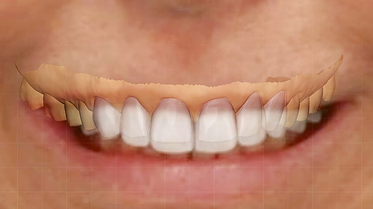
KOMPETENZ PLUSSeiten: 6-16, Sprache: DeutschFehmer, Vincent / Burkhardt, Felix / Sancho-Puchades, Manuel / Hämmerle, Christoph / Sailer, IrenaDie Diagnostik ist entscheidend für den vorhersagbaren Erfolg einer restaurativen Therapie. Hierbei müssen sich Patient und Zahnarzt vor der Anfertigung der definitiven Restauration auf ein gemeinsames Behandlungsziel einigen, um später Enttäuschungen zu vermeiden. Allerdings kann es schwierig sein, die Patientenwünsche vollständig zu erfassen. Ein nützliches Hilfsmittel zur Lösung dieses Problems sind das diagnostische Wax-up und Mock-up. Mit diesem zeitaufwendigen Verfahren wird jedoch nur eine einzige Variante des möglichen Behandlungsergebnisses visualisiert. Die moderne Digitaltechnik bietet nützliche Funktionen, die bei diesem diagnostischen Schritt zu einer Entscheidungsfindung beitragen können. Im vorliegende Beitrag werden die Möglichkeiten der Digitaltechnik für die prothetische Diagnostik erörtert und die Verfahren anhand von Patientenfällen beschrieben.
QZ - Quintessenz Zahntechnik, 1/2021
Exzellente Dentale ÄsthetikSeiten: 7-8, Sprache: DeutschPiskin, Cem / Burkhardt, FelixThe International Journal of Prosthodontics, 5/2020
DOI: 10.11607/ijp.6778, PubMed-ID: 32956436Seiten: 546-553, Sprache: EnglischPitta, João / Bijelic-Donova, Jasmina / Burkhardt, Felix / Fehmer, Vincent / Närhi, Timo / Sailer, IrenaPurpose: To evaluate the effect of cementation protocols on the bonding interface stability and pull-out forces of temporary implant-supported crowns bonded on a titanium base abutment (TiB) or on a temporary titanium abutment (TiA).
Materials and Methods: A total of 60 implants were restored with PMMA-based CAD/CAM crowns. Five groups (n = 12) were created: Group 1 = TiB/SRc: crown conditioned with MMA-based liquid (SR Connect, Ivoclar Vivadent); Group 2 = TiB/50Al-MB: crown airborne particle–abraded with 50-μm Al2O3 and silanized (Monobond Plus, Ivoclar Vivadent); Group 3 = TiB/30SiOAl-SRc: crown airborne particle–abraded with 30-μm silica-coated Al2O3 (CoJet, 3M ESPE) and conditioned with MMA-based liquid (SR Connect); Group 4 = TiB/30SiOAl-MB: crown airborne particle–abraded with 30- μm silica-coated Al2O3 (CoJet) and silanized (Monobond Plus); and Group 5 = TiA/TA-PMMA: crown manually enlarged, activated, and rebased with PMMA resin (Telio Lab, Ivoclar Vivadent). Specimens in the TiB groups were cemented using a resin cement (Multilink Hybrid Abutment, Ivoclar Vivadent). After aging (120,000 cycles, 49 N, 1.67 Hz, 5°C to 55°C, 120 seconds), bonding interface failure was analyzed (50x). Pull-out forces (N) (0.5 mm/minute) and modes of failure were registered. Chi-square and Kruskal-Wallis tests were used to analyze the data (α = .05).
Results: Bonding failure after aging varied from 0% (Group 5) to 100% (Groups 1, 2, and 4) (P .001). Mean pull-out force ranged between 53.1 N (Group 1) and 1,146.5 N (Group 5). The pull-off forces were significantly greater for Group 5 (P .05), followed by Group 3 (P .05), whereas the differences among the remaining groups were not significant (P > .05).
Conclusion: The cementation protocol had an effect on the bonding interface stability and pull-out forces of PMMA-based crowns bonded on a titanium base. Airborne particle abrasion of the crown internal surface and conditioning it with an MMA-based liquid may be recommended to improve retention of titanium base temporary restorations. Yet, for optimal outcomes, conventional temporary abutments might be preferred.
QZ - Quintessenz Zahntechnik, 5/2020
ErfahrungsberichtSeiten: 510-527, Sprache: DeutschFehmer, Vincent / Burkhardt, Felix / Sancho-Puchades, Manuel / Hämmerle, Christoph / Sailer, IrenaDie Diagnostik ist entscheidend für den vorhersagbaren Erfolg einer restaurativen Therapie. Hierbei müssen sich Patient und Zahnarzt vor der Anfertigung der definitiven Restauration auf ein gemeinsames Behandlungsziel einigen, um später Enttäuschungen zu vermeiden. Allerdings kann es schwierig sein, die Patientenwünsche vollständig zu erfassen. Ein nützliches Hilfsmittel zur Lösung dieses Problems sind das diagnostische Wax-up und Mock-up. Mit diesem zeitaufwendigen Verfahren wird jedoch nur eine einzige Variante des möglichen Behandlungsergebnisses visualisiert. Die moderne Digitaltechnik bietet nützliche Funktionen, die bei diesem diagnostischen Schritt zu einer Entscheidungsfindung beitragen können. Im vorliegende Beitrag werden die Möglichkeiten der Digitaltechnik für die prothetische Diagnostik erörtert und die Verfahren anhand von Patientenfällen beschrieben.
Schlagwörter: CAD/CAM, Mock-up, Visualisierung, additive Fertigung, 3-D-Druck
The International Journal of Prosthodontics, 4/2020
DOI: 10.11607/ijp.6402, PubMed-ID: 32639697Seiten: 380-385, Sprache: EnglischLee, Hyeonjong / Burkhardt, Felix / Fehmer, Vincent / Sailer, IrenaPurpose: To test the accuracies of different methods of digital vertical dimension augmentation (VDA) by comparison with a clinical situation.
Materials and Methods: Bite registrations with approximately 5 mm of VDA were made in the incisor regions of 10 subjects (mean VDA 4.5 mm). The conventional maxillary and mandibular stone casts in maximum intercuspation (MICP) and VDA bite registrations were digitized for all subjects using a laboratory scanner (control group). Lateral portraits were taken of all subjects to locate the position of the condylar axis. Four different digital VDA methods were compared to the control group: 100% rotation of the mandible referring to the lateral picture (100RL); 85% rotation and 15% translation referring to the lateral picture (85R15TL); 100% rotation in normal mounting mode of the Trios virtual articulator (100R); and jaw-motion analysis (JMA) equipment. The amount of VDA for each experimental group was compared to the control group. The augmented distances between the central incisors and the second molars were measured using 3D analyzing software. The ratio of the augmented distances between the posterior and anterior regions (P/A ratio) was calculated. One-way analysis of variance and multiple comparisons via least significant difference test were carried out to determine statistical significance.
Results: The P/A ratio of each group was as follows: Control = 0.61; 100RL = 0.55; 85R15TL = 0.61; 100R = 0.53; JMA = 0.52. Significant differences were observed for control vs JMA and for 85R15TL vs JMA (P .05). The addition of translational movement was the primary factor for increasing the accuracy of digital VDA, with the lateral picture being a secondary factor.
Conclusion: VDA using a virtual articulator with 100% rotation induces an error when compared to the clinical situation. When a clinician performs digital VDA, the setting of 85% rotation and 15% translation produces results closer to the real clinical condition.
International Journal of Computerized Dentistry, 1/2020
ApplicationPubMed-ID: 32207463Seiten: 73-82, Sprache: Deutsch, EnglischBurkhardt, Felix / Strietzel, Frank Peter / Bitter, Kerstin / Spies, Benedikt ChristopherHintergrund: Für den Behandlungserfolg einer implantatprothetischen Versorgung ist die Positionierung der Implantate im Knochen von hoher Bedeutung. Insbesondere bei einteiligen Implantaten ist eine nachträgliche Korrektur der Implantatachse nur erschwert möglich. Eine prothetisch orientierte, digitale Planung in Kombination mit einer vollgeführten Implantation kann in diesem Fall ein erhöhtes Maß an Sicherheit bieten.
Fallpräsentation: Zur Rehabilitation einer weitspannigen Schaltlücke in Regio 13 bis 16 wurden drei einteilige Implantate aus Zirkonoxid zur Aufnahme einer Zirkonoxidbrücke mit Extensionsbrückenglied geplant. Für eine optimale Positionierung basierte die Planung auf einem digitalen Set-up und dreidimensionalen DICOM-Daten (Digital Imaging and Communications in Medicine). Die geführte Implantatinsertion wurde mithilfe von Aufsätzen am chirurgischen Winkelstück sowie einer dazu kompatiblen, hülsenlosen Bohrschablone durchgeführt. Für die definitive Versorgung erfolgte die Abformung der Implantate mittels Intraoralscanner. Hierbei auftretende reflexionsbedingte Ungenauigkeiten wurden durch eine digitale Abutmentgeometrie ausgeglichen. Auf Basis dieser überlagerten STL-Daten wurde eine monolithische Zirkonoxid-Restauration subtraktiv gefertigt und adhäsiv zementiert.
Schlussfolgerung: Aufgrund einer vorausschauenden, prothetisch orientierten Planung konnten die einteiligen Keramikimplantate auch ohne Guide-Lösung des Implantatherstellers inseriert werden. Dies war mithilfe von Aufsätzen am chirurgischen Winkelstück und einer dazu kompatiblen Bohrschablone möglich. Dies ist besonders bei einteiligen Implantaten empfehlenswert, da eine nachträgliche Korrektur der Implantatachse nur durch irreversibles Beschleifen der Abutments möglich ist. Durch die Überlagerung der Scandaten mit der importierten, idealen Abutementgeometrie konnte die Genauigkeit der Implantatabformung verbessert werden. Es konnte gezeigt werden, dass mithilfe dieser Methode zukünftig auf eine Gingivaretraktion bei der Abformung dieses Implantattyps verzichtet werden kann.
Schlagwörter: Keramikimplantate, geführte Implantation, backward planning, Intraoralscan, CAD/CAM, digitaler Workflow
QZ - Quintessenz Zahntechnik, 7/2019
StatementSeiten: 906-907, Sprache: DeutschBurkhardt, Felix / Fehmer, VincentDentista, 2/2019
FokusSeiten: 21-23, Sprache: DeutschBurkhardt, Felix / Fehmer, Vincent / Sailer, Irena / Pjetursson, Bjarni E.Eine systematische Literaturübersicht


